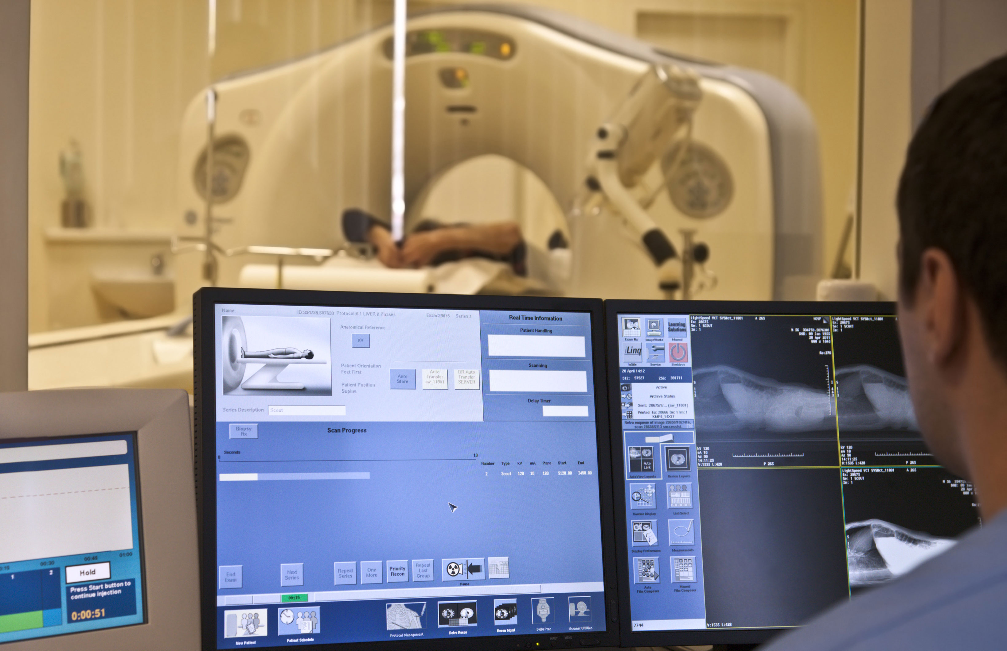
Beyond regular x-rays, MRI is the workhorse and mainstay imaging method for a large number of musculoskeletal conditions. It has been shown to be accurate and sensitive for the diagnosis of the most common injuries to the knee and shoulder. It is also indispensible in the investigation of back injuries. Magnetic resonance imaging is so good at showing changes in soft tissue, as well as bone, that it is often all that is needed to make a diagnosis. This has led to a dramatic increase in utilization over the past couple of decades, not all of it to the benefit of the person being imaged. Between 1995 and 2005 there was a 300% increase in MRI imaging of the low back without a measurable increase in positive patient outcomes.
The availability of MRI and the expected easy answer to a patient’s problem has led too many clinicians to forget or neglect their history taking and physical examination skills. In one study of work related shoulder injuries approximately half of the initial requests for MRIs from general practitioners did not have documented physical findings to support the request. As mentioned in a previous newsletter, the lack of documentation has many reasons beyond MRI’s promise of a quick diagnosis and while some of these reasons are understandable, they cannot all be excusable.
The problem is compounded when the issue of causation gets introduced. MRIs of the low back, neck, knee, shoulder, etc. can show us a great many things, including degenerative changes, previous injuries and co-morbid conditions, as well as increased signal in tendons, bursa, surrounding soft tissue and bone of questionable significance. If all goes well for the patient, the treating healthcare provider can put the information gained from the MRI together with the history and physical exam to arrive at an accurate diagnosis and successful treatment plan. Very often the determination of a diagnosis and initiation of treatment can be, and is, done without a determination of causation, sometimes without any thought about it at all.
Causation is typically, or at least should be, thought of early in work related cases since testing and treatment are dependent on establishing that the injury or condition happened as a result of the patient’s job. Personal injury cases can be more problematic. In many cases, the treating provider does not need to know the cause of the problem to treat it. The orthopedic surgeon doesn’t have to know if a meniscus tear was caused by an injury at home, at work or at a grocery store to do a meniscectomy, likewise for the neurosurgeon and a herniated disk. Human nature being as it is, we often do what we have to, e.g. the minimum amount of paperwork and time to make the diagnosis and treat the patient, rather than what we should do, e.g. document a thorough history and physical exam that can support the diagnosis, necessity of treatment and causation (if possible).
The question often arises as to whether an MRI can help in the determination of causation. Very frequently it can. There are very well established patterns of MRI findings in the knee for a number of mechanisms of injury – pivot shift, dash board, patellar dislocation, hyperextension, clipping. These patterns show a relationship between the mechanism of injury and cartilage damage, ligament tears and importantly bone contusions. These findings will be seen on MRI in the acute and subacute phases of injury but the bone contusions will resolve over time. This can be extremely helpful in determining a temporal relationship between injury and mechanism as well as plausibility. This method is less than foolproof however, since there is a paucity of research on the exact timeline of bone contusion/bone marrow edema resolution. Initial literature seemed to indicate that it resolved in 6-12 weeks. There is newer evidence based on longitudinal studies that show that most will be resolved by 42 weeks, but some can still be present over one year.
The literature is even less helpful in cases involving the shoulder and spine. There may be some findings that would help support an acute traumatic rotator cuff tear, for example, and there is fairly extensive literature on the MRI findings in chronic cuff tears. Unfortunately, there is a large gray zone in between where MRI can show the presence of a tear but give little if any information on the timing of the tear. Current treatment guidelines for the shoulder do not recommend early MRI scanning without clear cut physical exam findings of cuff dysfunction. It is typical that a patient undergo weeks or months of conservative treatment before imaging is ordered. The same is true for low back injuries. In the absence of distinct neurologic deficits early imaging is discouraged. By the time a MRI is obtained any evidence of the acute injury may have resolved or be indeterminate.
Treating medical providers are usually less than enthusiastic about providing reports of causation to begin with, let alone when there may be little objective evidence, from their own records or imaging reports, and even less literature to support it. The medical literature is primarily focused on diagnostic accuracy and outcomes. The medical community, which includes insurance providers, is also concerned with cost containment These topics push the issue of causation to the wayside and many patients get pushed along with it.
